 English
 Turkish
 Deutsch



What Diagnostic Imaging Is Used for Wisdom Tooth Removal?
Radiographic imaging is used when it is expected to contribute clinically relevant information to diagnosis or treatment planning. Panoramic radiography is commonly used for third-molar assessment because it provides a broad two-dimensional view of tooth position, root morphology, surrounding bone, adjacent teeth, and nearby anatomical structures.
However, panoramic radiography is not required in exactly the same way for every patient, and the choice of imaging should be based on the clinical situation.
For mandibular third molars, certain panoramic features may suggest a close relationship between the roots and the inferior alveolar canal. When additional three-dimensional information could influence the treatment plan, limited-field CBCT may be considered.
CBCT should not be used routinely for every impacted wisdom tooth. Its use should be justified by the expected diagnostic or surgical benefit.
Important radiographic considerations may include:
- Inferior Alveolar Canal Relationship: Evaluating the relationship between mandibular third-molar roots and the inferior alveolar canal when relevant to surgical planning.
- Root Morphology: Assessing root number, curvature, divergence, fusion, and other features that may influence extraction.
- Maxillary Sinus Relationship: Evaluating the relationship between maxillary third-molar roots and the sinus when clinically relevant.
- Adjacent Second Molar: Assessing caries, periodontal bone loss, root resorption, or other damage involving the neighboring tooth.
- Associated Pathology: Identifying cystic changes, enlarged follicular spaces, or other abnormalities requiring further assessment.
What Anesthesia Options Are Available for Removal Procedures?
The choice of anesthesia for wisdom tooth removal depends on procedural complexity, patient age, anxiety level, medical history, number of teeth being treated, expected surgical duration, clinical setting, and provider qualifications.
Local anaesthesia is commonly used to provide pain control at the surgical site and is sufficient for many straightforward and surgical third-molar removals.
Sedation may be considered for selected patients who have significant anxiety, limited tolerance for the procedure, or more extensive treatment needs. The depth of sedation should be described according to the actual level of consciousness rather than the route used to administer medication.
Moderate sedation allows purposeful response to verbal commands or light tactile stimulation, with spontaneous ventilation generally maintained. Deep sedation produces a deeper level of consciousness in which airway or ventilatory support may occasionally be required.
General anaesthesia produces a state of unconsciousness and may be appropriate for selected complex cases or patients when clinically justified and performed in an appropriately equipped setting by qualified personnel.
Anesthesia selection should be individualized and should not be determined solely by whether the tooth is described as impacted.
What Is the Step-by-Step Surgical Process for Tooth Removal?

The surgical approach to wisdom tooth removal is tailored to the position of the tooth, degree of impaction, root anatomy, surrounding bone, and proximity to important structures. After the selected anaesthetic technique is administered and adequate pain control is confirmed, the clinician creates surgical access when necessary.
For impacted teeth, a mucoperiosteal flap may be reflected to expose the underlying bone. Bone removal is performed only to the extent required for controlled access to the tooth.
The tooth may then be sectioned into smaller components when this facilitates safer and more conservative removal. Sectioning is not required in every case and depends on tooth position, root morphology, and surgical access.
A typical surgical sequence may include:
- Surgical Access: Creating an incision and reflecting a flap when necessary to expose the operative site.
- Bone Removal: Removing the minimum amount of surrounding bone required to access and deliver the tooth safely.
- Tooth Sectioning: Dividing the crown or roots when sectioning reduces the amount of bone removal or force required for extraction.
- Socket Assessment: Inspecting and irrigating the extraction site and removing pathological tissue when clinically indicated.
- Surgical Closure: Repositioning the soft tissues and placing sutures when required.
The exact sequence varies according to the individual surgical situation, and not every wisdom tooth removal requires all of these steps.
What Postoperative Symptoms Are Normal After Tooth Removal?
Temporary postoperative symptoms are common following third molar surgery and generally reflect the inflammatory response and tissue trauma associated with the procedure.
Swelling often increases during the first few postoperative days before gradually improving. Mild bruising, restricted mouth opening, blood-tinged saliva, and localized discomfort may also occur. The severity and duration of these symptoms vary according to surgical complexity, number of teeth removed, patient factors, and individual healing response.
Common temporary postoperative findings may include:
- Facial Swelling: Localized swelling can increase during the first two to three days and generally decreases gradually afterward.
- Jaw Stiffness (Trismus): Temporary limitation in mouth opening may occur because of inflammation and discomfort affecting the masticatory muscles.
- Mild Bleeding or Oozing: Small amounts of blood or blood-tinged saliva may occur during the early postoperative period.
- Localized Discomfort: Pain or tenderness around the extraction site is common and should generally improve over time with appropriate postoperative care.
Persistent or worsening symptoms should be assessed by the treating dental team.
What Is Alveolar Osteitis (Dry Socket) and How Is It Managed?
Alveolar osteitis, commonly called dry socket, is a painful postoperative complication that can occur after tooth extraction, particularly following mandibular third molar removal. It is associated with breakdown or loss of the blood clot and delayed organization of the extraction socket.
Patients typically develop increasing pain several days after extraction rather than the expected gradual improvement. The pain may radiate toward the ear, temple, or surrounding jaw region. An unpleasant taste or odor may also occur.
Dry socket is primarily managed by controlling symptoms while the socket heals naturally.
Management may include:
- Gentle Irrigation: The socket may be irrigated to remove accumulated debris without aggressive curettage.
- Local Dressing When Indicated: A medicated dressing may be placed for short-term pain relief in selected patients.
- Analgesic Management: Appropriate pain medication may be recommended according to the patient’s medical history and symptom severity.
- Oral Hygiene Instructions: Patients may receive specific instructions for keeping the area clean while avoiding trauma to the healing socket.
- Clinical Review: Follow-up may be required if pain persists or symptoms do not improve as expected.
Dry socket is not the same as a spreading bacterial infection and does not routinely require systemic antibiotics unless there is a separate clinical indication.
How Long Does Recovery Take After Wisdom Tooth Removal?
Recovery after wisdom tooth removal varies according to impaction depth, surgical difficulty, number of teeth removed, patient age, general health, smoking status, and individual healing response.
Swelling, discomfort, and limited mouth opening often improve substantially during the first several days, but soft-tissue healing continues beyond the initial symptomatic recovery period.
Bone formation within the extraction socket develops progressively over the following weeks and months, while remodeling continues for a longer period.
Following individualized recovery after extraction protocols can support appropriate healing and help patients identify complications early.
Many patients are able to return gradually to routine daily activities within several days, but there is no universal recovery timeline. Strenuous exercise and activities that increase bleeding risk may need to be limited temporarily according to the surgeon’s instructions.
How Much Does Wisdom Tooth Removal Cost Across Countries?
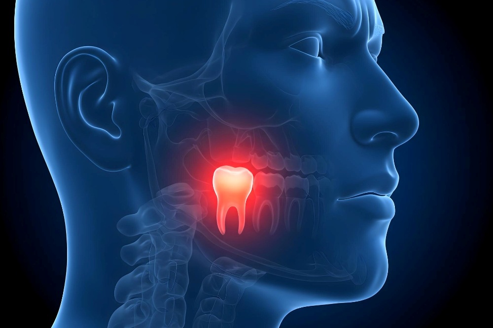
The cost of wisdom tooth removal varies according to the degree of impaction, surgical complexity, anaesthesia requirements, imaging, clinician expertise, facility, geographic location, and whether additional procedures are required.
A straightforward erupted third molar is generally less complex than a deeply impacted tooth requiring flap elevation, bone removal, or sectioning, but fees vary considerably between clinics and healthcare systems.
Treatment in Turkey may be less expensive than private third molar surgery in some other countries because of differences in operating expenses, labour costs, exchange rates, and healthcare market conditions. However, percentage savings cannot be guaranteed and should be calculated using current, directly comparable treatment quotations.
Patients should confirm whether quoted fees include consultation, radiographic imaging, CBCT when required, anaesthesia or sedation, medications, follow-up visits, and management of postoperative complications.
Current prices should therefore be confirmed directly with the treating clinic because costs can change over time and vary substantially between individual cases.
What Postoperative Care Supports Healing After Removal?
Postoperative care can support healing, reduce discomfort, and help lower the risk of certain complications, but no aftercare routine can guarantee an uncomplicated recovery.
Immediately after extraction, gauze pressure may be used when required to control bleeding and support initial clot formation. Patients should follow the specific instructions provided by their surgical team regarding how long to maintain pressure and when to replace or remove the gauze.
Cold packs may be recommended intermittently during the early postoperative period to help manage swelling and discomfort. The usefulness of heat later in recovery depends on the clinical situation and should follow the treating clinician’s advice.
Common postoperative recommendations may include:
- Follow Dietary Instructions: Soft foods may be more comfortable during the early recovery period. Progress toward a normal diet should be based on comfort and the surgeon’s recommendations.
- Protect the Surgical Site: Avoid smoking and other behaviors that may interfere with clot stability or wound healing.
- Maintain Appropriate Oral Hygiene: Clean the mouth according to the surgeon’s instructions while avoiding direct trauma to the extraction site during the early healing period.
- Use Rinses as Directed: Salt-water or prescribed rinses may be recommended at an appropriate stage after surgery, depending on the procedure and clinician’s protocol.
- Modify Activity Temporarily: Strenuous exercise or activities that may increase bleeding or trauma should be limited when advised.
- Take Medication as Directed: Use analgesics according to instructions. Antibiotics should only be used when prescribed for a specific clinical indication.
- Attend Follow-Up When Required: Seek review if pain, swelling, bleeding, sensory changes, fever, or other symptoms are persistent, worsening, or unexpected.
What Rare Complications Can Occur with Tooth Removal?
Third molar removal is a common oral surgical procedure, but complications can occur because of the close relationship between wisdom teeth and surrounding nerves, bone, soft tissues, and the maxillary sinus.
In mandibular third molar surgery, the inferior alveolar nerve and lingual nerve are important anatomical structures. Injury, compression, inflammation, or surgical trauma involving these nerves can lead to altered sensation.
Inferior alveolar nerve disturbance may affect sensation in the lower lip or chin, while lingual nerve disturbance can affect sensation in the tongue and, in some cases, taste perception. Many postoperative sensory disturbances improve over time, but persistent or permanent changes are possible.
In the maxilla, third molar roots may lie close to the maxillary sinus. Removal can occasionally create an oroantral communication between the extraction site and the sinus. Small communications may close with appropriate management, while larger or persistent defects may require surgical closure and specific postoperative sinus precautions.
Other uncommon complications can include infection, excessive bleeding, displacement of tooth or root fragments into adjacent anatomical spaces, fracture of surrounding bone, or delayed healing. The likelihood of these complications depends on anatomy, surgical difficulty, patient health, and other clinical factors.
When Is Coronectomy an Alternative to Complete Tooth Removal?
Coronectomy is a surgical technique used in selected mandibular third molars when complete extraction is considered to carry an increased risk of inferior alveolar nerve injury.
The decision is based on clinical and radiographic assessment. CBCT may be used when three-dimensional imaging is clinically indicated to clarify the relationship between the tooth roots and the inferior alveolar canal, but coronectomy is not determined by CBCT findings alone.
During coronectomy, the crown of the third molar is removed while all or part of the roots are intentionally retained within the jaw. The remaining root structure is positioned sufficiently below the surrounding bone level to allow soft-tissue and bone healing, provided the roots are suitable for retention.
Coronectomy is generally considered only when the retained roots are not associated with conditions that would make intentional retention inappropriate, such as certain forms of infection, mobility, or other pathology.
Retained roots may remain stable or migrate over time. Root migration is relatively common and does not automatically represent treatment failure. A minority of cases may require later surgical intervention because of root exposure, symptoms, infection, or other complications.
In appropriately selected high-risk mandibular third molars, coronectomy can reduce the risk of inferior alveolar nerve injury compared with complete extraction, although it introduces its own potential complications and follow-up requirements.
What Questions Should You Ask Before Wisdom Tooth Removal?
A preoperative consultation should help patients understand why removal is being recommended, what anatomical risks are present, which treatment alternatives are available, what type of anaesthesia is planned, and what postoperative care will be required.
Important questions to discuss include:
- Clinical Indication: Why is removal recommended for this particular wisdom tooth, and is monitoring a reasonable alternative?
- Anatomical Relationship: How close is the tooth to the inferior alveolar nerve, lingual structures, adjacent teeth, or maxillary sinus?
- Imaging Requirements: Is additional imaging such as CBCT likely to change the surgical plan?
- Surgical Strategy: Is complete extraction appropriate, or should coronectomy or another approach be considered?
- Anaesthesia Plan: What level of anaesthesia or sedation is appropriate for my medical history, anxiety level, and surgical complexity?
- Potential Complications: What are the specific risks of nerve disturbance, dry socket, infection, sinus communication, bleeding, or other complications in my case?
- Recovery Expectations: What symptoms are expected, which activities should be limited, and which warning signs require urgent review?