Dental tomography is a radiological imaging technique performed by rotating a cone-shaped X-ray beam and an image detector simultaneously around the patient’s head, typically between 180 and 360 degrees. This rotation allows the selected volume to be viewed on three different planes (axial, sagittal, and coronal) using computer software.
What Is the Difference Between Dental Tomography and Conventional Dental X-rays?
The main difference between dental tomography and traditional dental X-rays is the ability to capture three-dimensional (3D) images. This 3D imaging provides a complete view of the teeth, jawbone, and airway in terms of length, width, and depth.
Who Needs Dental Tomography and When Is It Used?
Dental tomography is used for patients who experience jaw pain, lesions, trauma, or suspected fractures. It is also used for implant planning and removal, bone graft evaluations, impacted tooth assessments before surgery, jaw cysts and tumors, temporomandibular joint (TMJ) imaging, root canal treatments, and surgical planning for prosthetic restorations.
Advantages of Dental Tomography
Dental tomography provides a high-quality 3D perspective with excellent image resolution and diagnostic accuracy. It allows clear visualization of bone structures and enables more precise diagnosis and treatment planning.
3D Imaging: Offers detailed, layered images that improve treatment precision.
Accurate Diagnosis: Enhances the ability to identify and manage complex jaw disorders.
Improved Implant Planning: Ensures accurate implant positioning and sizing.
Quick Scanning with Low Radiation: Provides fast scanning with minimal radiation exposure, ensuring patient comfort.
Why Is Dental Tomography Important?
Dental tomography helps visualize the full shape and dimensions of the jawbone, allowing for successful implant surgeries. It identifies infections and their exact locations, and it can detect abnormal lesions that may indicate bone cancers, tumors, or cysts.
Does Dental Tomography Involve Radiation?
Dental tomography uses a lower dose of radiation compared to conventional dental X-rays. One of its biggest advantages is producing high-quality images with minimal radiation exposure. To illustrate, a standard chest X-ray exposes a patient to approximately 50 times more radiation than a dental tomography scan.
As implant treatments and TMJ disorders become increasingly common, the use of advanced imaging technologies is growing as well. With its high resolution and low-dose properties, dental tomography has revolutionized dentomaxillofacial radiology—offering a new path for diagnosis and treatment planning. It should be used particularly in cases where two-dimensional radiographs are insufficient and more detailed examination is required.
Frequently Asked Questions About Dental Tomography
Question 1: What is dental tomography?
Dental tomography is an imaging technique that allows dentists and oral surgeons to visualize the teeth and jaw in three dimensions. It is used to diagnose oral issues and assist in treatment planning.
Question 2: How does dental tomography work?
It uses a rotating X-ray source and detector that move around the patient’s head. The process captures multiple two-dimensional slices, which are then reconstructed into a 3D model of the oral and maxillofacial area.
Question 3: Who is suitable for dental tomography?
It is used for implant placement, oral or jaw surgery, root canal treatments, and TMJ disorder assessments. The necessity of the procedure is determined by a dentist or oral surgeon.
Question 4: How long does a dental tomography scan take?
The scan is usually very quick—typically completed within a few minutes depending on the required detail and number of images.
Question 5: Does dental tomography expose patients to radiation?
Yes, but the radiation dose is significantly lower than that of conventional computed tomography (CT) scans. The dose is optimized to minimize exposure while ensuring clear imaging results.
Question 6: Is the dental tomography procedure uncomfortable?
The procedure is generally painless and causes minimal discomfort. The patient simply needs to remain still and follow the technician’s instructions.
Question 7: How soon are the results available?
Results are typically reviewed immediately after the scan, and in most cases, can be discussed with the patient the same day.
Question 8: What are the main advantages of dental tomography?
Detailed and Accurate Imaging: Provides precise, multi-dimensional views of teeth and jaw structures.
Fewer Repeat Scans: Offers comprehensive images, reducing the need for multiple X-rays.
Fast and Convenient: Easier and quicker to perform compared to traditional radiography.
 English
 Turkish
 Deutsch



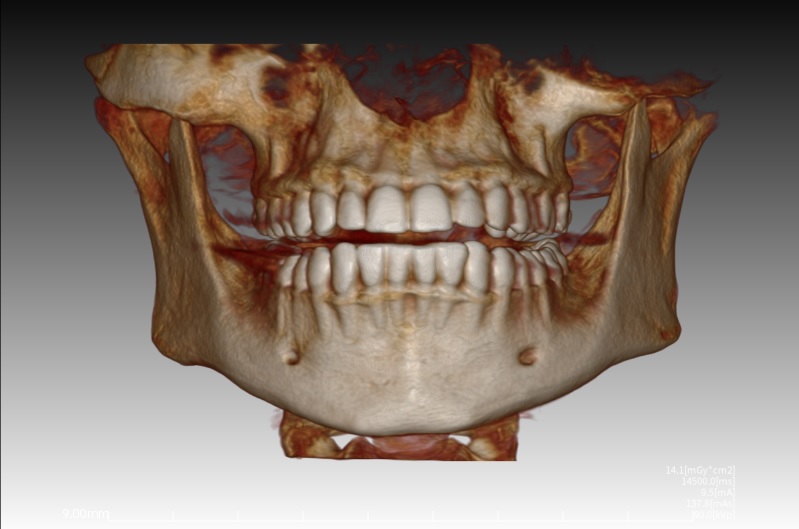 Dental tomography helps visualize the full shape and dimensions of the jawbone, allowing for successful implant surgeries. It identifies infections and their exact locations, and it can detect abnormal lesions that may indicate bone cancers, tumors, or cysts.