 English
 Turkish
 Deutsch


Dental X-rays are imaging techniques used by dentists to detect problems related to the teeth and gums. During a clinical examination, issues on the visible surfaces of the teeth or the exposed parts of the gums can be identified. However, it is not possible to see problems in the deeper parts of the gums, within the bone, or at the tooth roots with the naked eye. This is where medical imaging methods come into play. Different types of X-rays are used to visualise structures that cause health problems, making it possible to detect issues that cannot be seen during a standard clinical examination. In this article, we will discuss how dental X-rays are taken and whether pregnant women can safely undergo dental X-rays. First, let’s look at which imaging methods are commonly used.
What Are the Types of Dental X-rays?
In general, we can list the main types of dental X-rays as follows:
- Periapical Dental X-ray
This type of X-ray provides a complete image of the tooth, including the roots, and is therefore one of the most commonly used methods. When it is necessary to evaluate the structure of the tooth roots in particular, periapical X-rays are preferred. In addition to the roots, they also help assess bone levels and detect formations such as cysts and abscesses.
- Panoramic Dental X-ray
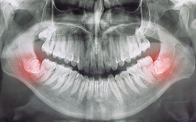
Sometimes it is necessary to visualise the entire oral structure to identify the underlying causes of a patient’s complaints. In such cases, a panoramic dental X-ray is used. With a single image, both the upper and lower teeth, as well as part of the jaw, can be evaluated. Panoramic X-rays are also used to assess dental development in children as well as adults. When patients attend for their first examination, it is often necessary to evaluate the overall condition of the mouth and teeth, and a panoramic X-ray is commonly taken for this purpose.
- Cephalometric Dental X-ray
This imaging method shows the skull bones and soft tissues from the front, back and side views. Cephalometric X-rays are frequently used in orthodontic treatments and in cases where orthognathic surgery is required.
- Dental CT (Dental Tomography)
Dental CT is an imaging method used to obtain a clear and detailed view of targeted structures in a specific region. It consists of thin slices and also includes volumetric information, and is taken using X-rays. Although it is often confused with conventional dental X-rays, there are significant differences between the two. The most important difference is that tomography provides 3D images. This makes it possible to accurately assess the length, depth and width of the area of interest. Dental CT can visualise not only all the teeth, but also the airway and jawbone.
Are Dental X-rays Harmful?
First, let’s clarify a common concern: whether dental X-rays are harmful. It is true that the X-rays used in radiography contain a certain amount of radiation. However, the amount of radiation used in modern dental X-ray systems is very low. Furthermore, today’s imaging devices are far more advanced than traditional machines and emit significantly less radiation during exposure.
Of course, the exact radiation dose varies depending on which device and which type of X-ray is used. To give a general idea, the radiation dose from a small, standard dental X-ray is around 0.001 mSv, and a panoramic radiograph is around 0.003 mSv. This is roughly equivalent to the amount of radiation absorbed during a 4–5 hour flight. In other words, it is similar to the “background radiation” we are exposed to in daily life. It is also important to remember that every possible precaution is taken during X-ray procedures to keep radiation exposure as low as reasonably achievable.
How Is a Dental X-ray Taken?
For a dental X-ray, the patient is first taken to the X-ray room. After the necessary protective measures are applied, the patient’s jaw is positioned correctly on the X-ray unit. Special supports are placed on both sides of the head to stabilise it. Once all settings and adjustments are made on the device, the scan begins. The unit rotates around the head, captures images and sends them to a computer, where the dentist can evaluate them in detail.
This entire process does not cause any pain or discomfort. None of the imaging procedures used in dentistry are painful; they do not cause aches or any unpleasant sensations for the patient.
What Is a Panoramic Dental X-ray?
A panoramic dental X-ray is an imaging method that allows both the teeth and the jawbone to be viewed with a single exposure. In a sense, it provides a two-dimensional “picture” of the mouth. While it is often thought to be used only for visualising the teeth, panoramic imaging actually shows the teeth together with the jawbone. It also allows the dentist to clearly see multiple bones in the head and neck region, as well as important anatomical structures.
Dental caries and gum diseases are among the most common oral health problems. Panoramic X-rays play a crucial role in accurately diagnosing these conditions. They are also used to detect impacted teeth and to identify problems in the jaw joints and sinuses. In addition, they are invaluable for evaluating and planning dental implant treatments, as well as for checking existing implants.
In some cases, various lesions or masses such as tumours can develop in the mouth. These may not always be visible during a clinical examination. In such situations, panoramic imaging helps to identify these formations. It is also used when there is suspicion of oral cancer or when anomalies are suspected in the tissues or jawbones. One of the main reasons panoramic X-rays are preferred over standard X-rays is that they allow the entire oral structure to be assessed in a single image.
In traditional imaging methods, X-rays pass through the teeth and are captured on film inside the mouth. In a panoramic X-ray, however, the X-rays pass through the entire oral region and are detected by a digital sensor. Despite providing such detailed information, the procedure itself only takes a few seconds. The device rotates 180 degrees around the head, records a complete image of the mouth and transfers it to a computer. During the scan, it is sufficient for the patient to keep their head still and place their tongue against the roof of the mouth. As no pain, discomfort or negative sensations are experienced, having a panoramic X-ray is a very comfortable process for the patient.
Can Pregnant Women Have Dental X-rays?

As mentioned earlier, the radiation dose emitted during a dental X-ray is very low. Throughout the day, similar levels of radiation are emitted from sunlight, microwave ovens and communication devices. For this reason, it is difficult to argue that the radiation from dental X-rays will clearly harm the expectant mother or the baby in the womb.
However, even if the amount is extremely low, there is still some degree of radiation exposure. Therefore, it would not be accurate to claim that dental X-rays are completely risk-free during pregnancy. Expectant mothers should always consult their obstetrician or gynaecologist responsible for their prenatal care in order to receive more detailed and personalised information. If you have questions about dental X-rays, you can share them in the comments section.


